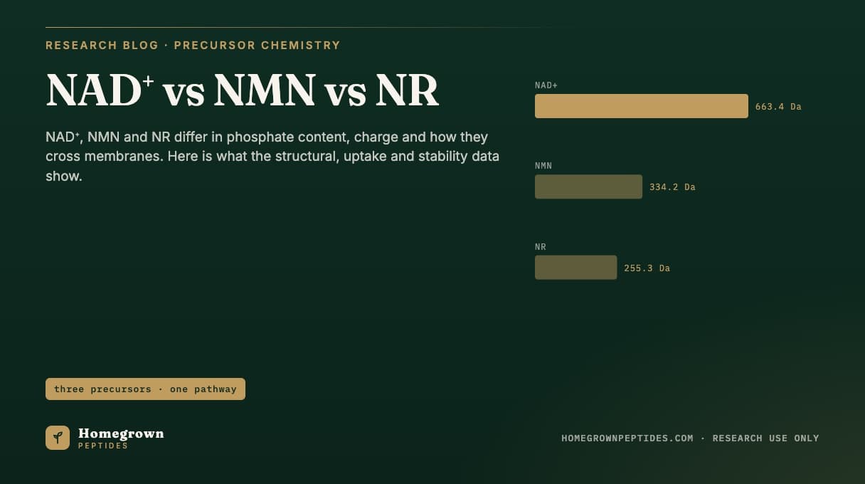
NAD+, NMN and NR are three distinct molecules in the same salvage pathway, not interchangeable names for one compound. NAD+ (C21H27N7O14P2, 663.43 g/mol) is the dinucleotide itself, two nucleotides joined through a pyrophosphate bridge. NMN (C11H15N2O8P, 334.22 g/mol) is one half of that molecule, nicotinamide attached to ribose-5-phosphate, one enzymatic step short of NAD+. NR (255.25 g/mol as the free cation, commonly supplied as a 291.71 g/mol chloride salt) is the same nicotinamide-ribose pair with the phosphate removed. They differ in phosphate content, net charge, and which route gets them into a cell: NR behaves like a membrane-permeant nucleoside, NMN requires either a disputed direct transporter or extracellular dephosphorylation to NR by CD73, and both are rebuilt to NAD+ intracellularly by the NRK and NMNAT enzyme pairs.
| Attribute | Detail |
|---|---|
| Chemical relationship | NAD+ = nicotinamide ribonucleotide (NMN) linked by pyrophosphate to adenosine monophosphate; NR = NMN minus its phosphate group |
| Rate-limiting salvage enzymes | NRK1/NRK2 phosphorylate NR to NMN; NMNAT1/2/3 adenylylate NMN to NAD+ (Ratajczak et al., 2016) |
| Extracellular NMN handling | Ecto-5'-nucleotidase CD73 dephosphorylates extracellular NMN to NR before uptake in cultured tumor cell models (Grozio et al., 2013) |
| Disputed direct NMN transporter | Slc12a8 reported to transport intact NMN across the plasma membrane in intestinal and other tissue models (Grozio et al., 2019) |
| Tissue distribution after dosing | Isotope-labeled precursors are extensively broken down to nicotinamide before resynthesis into NAD+ in mouse tissue flux studies (Liu et al., 2018) |
| Detection method | LC-MS/MS targeted metabolomics distinguishes NAD+, NMN, NR, NAM and NAAD by exact mass and retention time (Trammell and Brenner, 2013) |
How do NAD+, NMN and NR differ chemically?
All three share the same pyridine-ring nicotinamide group, the feature that gives NAD+ its redox chemistry. They differ in what is attached to that ring. NR is nicotinamide joined through an N-glycosidic bond to a ribose sugar, with no phosphate. Because the glycosidic bond quaternizes the pyridine nitrogen, NR is a permanent cation even before any phosphate is added, which is why it is typically supplied and stabilized as a chloride or other anion salt rather than a free base. NMN is NR with a phosphate group esterified to the 5' position of the ribose, adding a second, independently ionizable acidic group. NAD+ is NMN joined through a pyrophosphate linkage to a second nucleotide, adenosine monophosphate, giving the dinucleotide its two phosphate groups, its adenine ring, and its 663.43 g/mol mass. Each addition changes the molecule's polarity and net charge, the main reason the three are not handled identically by cell membranes (Bogan and Brenner, 2008).
Which enzymes convert each precursor toward NAD+?
The salvage route runs in one direction: NR to NMN to NAD+. Nicotinamide riboside kinases, NRK1 and the more tissue-restricted NRK2, phosphorylate NR to NMN using ATP. Nicotinamide mononucleotide adenylyltransferases, NMNAT1, NMNAT2 and NMNAT3, then join NMN to AMP to form NAD+, each isoform localized to a different subcellular compartment (nuclear, Golgi-associated and mitochondrial, respectively). Knockout and knockdown experiments in cultured cells and mice place NRK1 as the dominant rate-limiting step for both NR and, indirectly, NMN that first needs conversion to NR, since NRK1-null cells lose most of their response to extracellular NR and NMN alike (Ratajczak et al., 2016). NMN can also be generated directly from nicotinamide and phosphoribosyl pyrophosphate by NAMPT, the pathway cells use for intracellular NAD+ salvage independent of any externally supplied NMN or NR.
| Property | NAD+ | NMN | NR |
|---|---|---|---|
| Molecular formula | C21H27N7O14P2 | C11H15N2O8P | C11H15N2O5+ (cation) |
| Molecular weight | 663.43 g/mol | 334.22 g/mol | 255.25 g/mol (cation); 291.71 g/mol as chloride salt |
| CAS number | 53-84-9 | 1094-61-7 | 23111-00-4 (chloride salt) |
| Phosphate groups | 2 | 1 | 0 |
| Net charge at physiological pH | Approximately -1 (two ionized phosphates offset by the pyridinium nitrogen) | Approximately -1 (one ionized phosphate offset by the pyridinium nitrogen) | +1 (permanent pyridinium cation, no ionizable phosphate) |
| Membrane permeability evidence | Not membrane-permeant intact; extracellular NAD+ is degraded by CD38/CD73 before any component is taken up (Grozio et al., 2013) | Disputed; CD73-mediated dephosphorylation to NR is the consistently replicated route, direct Slc12a8 transport is contested (Grozio et al., 2013; 2019) | Taken up largely intact via nucleoside transport, then rephosphorylated intracellularly (Ratajczak et al., 2016; Trammell et al., 2016) |
| Key entry enzyme | NMNAT1/2/3 (forms NAD+ from NMN + AMP) | NAMPT (forms NMN from NAM); NRK1/2 (forms NMN from NR) | NRK1/NRK2 (forms NMN from NR) |
What did isotope-tracing studies in mice find about which form actually reaches tissues?
A comprehensive stable-isotope flux study measured NAD+ synthesis and breakdown across multiple mouse tissues using labeled nicotinamide and labeled NAD+ precursors tracked by LC-MS. A large fraction of circulating and tissue NAD+ turnover ran through nicotinamide as a common intermediate rather than through intact incorporation of whatever precursor was introduced: nicotinamide equivalents generated by extracellular and intracellular breakdown of NAD+, NMN and NR were recaptured and resynthesized into NAD+ by the NAMPT salvage step in most tissues examined (Liu et al., 2018). A separate metabolic profiling study compared nicotinamide, nicotinic acid, NR and tryptophan as starting precursors across mouse liver, kidney, heart and brain, and found that tissue-level expression of NRK1/2, NAPRT and NAMPT, not the precursor's identity alone, determines how efficiently a tissue converts a precursor to NAD+, with cardiac and skeletal muscle showing the highest NRK2-dependent use of the NR route (Mori et al., 2014). Neither study supports a model in which NMN or NR crosses from blood into tissue and is adenylylated to NAD+ without first passing through, or being broken down to, smaller intermediates.
What have in vitro uptake experiments shown, and is there a dedicated NMN transporter?
Working with cultured tumor cell lines treated with the NAMPT inhibitor FK866 to isolate precursor-dependent NAD+ recovery, one study showed that exogenous NMN added to the culture medium is hydrolyzed to NR by the ecto-enzyme CD73 (5'-nucleotidase) at the cell surface before the resulting NR is taken up and rephosphorylated intracellularly back to NMN; blocking CD73 activity blunted the ability of extracellular NMN to restore intracellular NAD+ in these cells (Grozio et al., 2013). A later study in cell lines and knockout mice reported that NRK1 is required for cells to use either extracellular NR or extracellular NMN efficiently, consistent with NMN needing conversion to NR, or needing an independent transporter, before NRK-mediated rephosphorylation can occur (Ratajczak et al., 2016). A separate report proposed Slc12a8 as a plasma-membrane transporter capable of moving intact NMN into cells, based on heterologous expression and isotope-labeled NMN uptake assays in small intestinal tissue from wild-type versus Slc12a8-knockout mice (Grozio et al., 2019). The two findings are not fully reconciled: CD73-mediated dephosphorylation has been independently replicated in multiple cell systems, while direct membrane transport of intact NMN through Slc12a8 remains an active point of discussion. Practically, an experiment adding NMN to cultured cells or an animal model is not necessarily delivering intact NMN across the membrane; depending on the system, ecto-nucleotidase activity at the cell surface may be the actual point of entry into the salvage pathway.
How stable is each precursor in aqueous solution and in storage?
NR's quaternary pyridinium nitrogen makes the N-glycosidic bond to its ribose susceptible to hydrolysis, and published chemical evaluations of NAD+ precursor vitamins describe this hydrolysis, which releases free nicotinamide, as pH- and temperature-dependent, proceeding faster as solution pH rises from acidic toward neutral (Bogan and Brenner, 2008). NMN carries the same nicotinamide-ribose linkage plus a labile phosphoester bond, giving it two possible degradation routes to nicotinamide: glycosidic hydrolysis and dephosphorylation. Building a targeted LC-MS/MS method for NAD+ metabolites required explicit attention to this instability, since method development noted that NMN and NR can continue converting to nicotinamide during sample handling if extraction is not fast enough to quench ongoing turnover, which would otherwise understate the intact precursor and overstate nicotinamide (Trammell and Brenner, 2013). NAD+ is comparatively more stable at its glycosidic bond, but remains subject to enzymatic degradation by NAD+ glycohydrolases such as CD38 in biological matrices rather than purified buffer.
How is each precursor detected and measured analytically?
NAD+, NMN, NR, nicotinamide (NAM) and the intermediate nicotinic acid adenine dinucleotide (NAAD) can be resolved from one another in a single LC-MS/MS run because each has a distinct exact mass and retention time on reverse-phase or ion-pairing chromatography; a dedicated targeted metabolomics method built around this separation reported detection of all five species from one tissue or cell extract using multiple-reaction-monitoring mass spectrometry (Trammell and Brenner, 2013). In mice and in humans dosed orally with NR, the same kind of LC-MS/MS panel tracked the full time course of NR, NAM, NAAD and NAD+ in blood, which is how researchers identified NAAD, rather than NAD+ itself, as the most sensitive circulating marker of increased NAD+ synthesis flux after an oral NR dose (Trammell et al., 2016). This panel approach, rather than a single-analyte assay, is standard in the literature because conversion between species happens fast enough that measuring only one misses most of what is occurring.
Background on NAD+ itself, including its two redox states and sirtuin-linked signaling roles, is covered in what is NAD+, and the distinction between NAD+ as a bound coenzyme versus a consumed substrate is addressed in is NAD+ a coenzyme or a substrate. The same mass-spectrometry identity logic applied here is covered generally in mass spectrometry for peptide identity, and the lyophilized-powder, cold-chain storage principles that limit the degradation chemistry above are covered in peptide storage and stability. Lot-specific identity and purity data for research-grade NAD+ are posted on our NAD+ lot page.
Frequently asked questions
Is NMN the same molecule as NAD+?
No. NMN is one of the two nucleotide halves that make up NAD+. NAD+ is formed when NMN is joined to AMP by an NMNAT enzyme, adding a second ring system and a second phosphate.
Is NR the same molecule as NMN?
No. NR is NMN without the phosphate group. An NRK enzyme adds that phosphate back, converting NR to NMN inside the cell.
Why do NMN and NR both end up measured as nicotinamide in some studies?
Both molecules contain a hydrolyzable glycosidic bond to nicotinamide, and NMN has an additional labile phosphoester bond. Isotope-tracing and LC-MS/MS studies have measured substantial conversion of both to free nicotinamide, in solution, during sample handling, and in cell and animal models, before any resynthesis into NAD+.
Does a research model need a specific transporter to use NMN?
The published literature has not settled this question. One line of evidence supports a direct plasma-membrane transporter, Slc12a8. A separate, independently replicated line of evidence shows extracellular NMN being dephosphorylated to NR by CD73 before cellular uptake. Both mechanisms have been reported in different cell and tissue systems.
Which precursor is most stable for long-term storage as a lyophilized powder?
The degradation chemistry discussed in the literature, glycosidic bond hydrolysis for NR and both glycosidic and phosphoester hydrolysis for NMN, applies to aqueous solution. Published stability evaluations of these NAD+ precursor vitamins describe the dry, lyophilized state as substantially slower to degrade than reconstituted solution for this class of molecule (Bogan and Brenner, 2008).
What models were these uptake and transporter studies conducted in?
Mouse tissue, mouse knockout models, and cultured human or rodent cell lines. This article describes laboratory research models; it is not a summary of clinical evidence and does not describe a protocol for use in any subject.
References
- Bogan KL, Brenner C. Nicotinic acid, nicotinamide, and nicotinamide riboside: a molecular evaluation of NAD+ precursor vitamins in human nutrition. Annu Rev Nutr. 2008;28:115-130. PubMed 18429699.
- Trammell SA, Brenner C. Targeted, LCMS-based metabolomics for quantitative measurement of NAD+ metabolites. Comput Struct Biotechnol J. 2013;4:e201301012. PubMed 24688693.
- Grozio A, Sociali G, Sturla L, et al. CD73 protein as a source of extracellular precursors for sustained NAD+ biosynthesis in FK866-treated tumor cells. J Biol Chem. 2013;288(36):25938-25949. PubMed 23880765.
- Mori V, Amici A, Mazzola F, et al. Metabolic profiling of alternative NAD biosynthetic routes in mouse tissues. PLoS One. 2014;9(11):e113939. PubMed 25423279.
- Trammell SA, Schmidt MS, Weidemann BJ, et al. Nicotinamide riboside is uniquely and orally bioavailable in mice and humans. Nat Commun. 2016;7:12948. PubMed 27721479.
- Ratajczak J, Joffraud M, Trammell SA, et al. NRK1 controls nicotinamide mononucleotide and nicotinamide riboside metabolism in mammalian cells. Nat Commun. 2016;7:13103. PubMed 27725675.
- Liu L, Su X, Quinn WJ 3rd, et al. Quantitative analysis of NAD synthesis-breakdown fluxes. Cell Metab. 2018;27(5):1067-1080.e5. PubMed 29685734.
- Grozio A, Mills KF, Yoshino J, et al. Slc12a8 is a nicotinamide mononucleotide transporter. Nat Metab. 2019;1(1):47-57. PubMed 31131364.
Research use only. Homegrown Peptides products are for laboratory research and are not for human or animal use. Nothing in this article is medical advice.