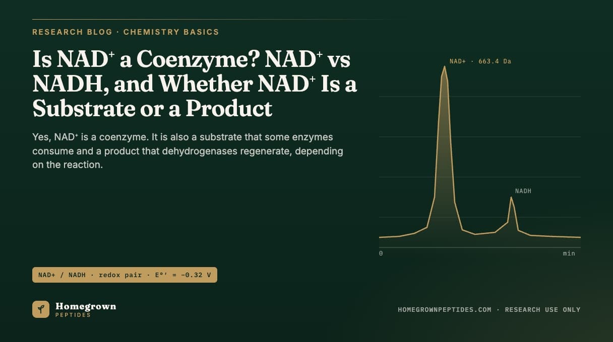
Yes, NAD+ is a coenzyme. It carries electrons, binding reversibly to enzyme active sites to accept or donate a hydride ion rather than becoming part of a permanent protein structure. NAD+ is also a substrate: dehydrogenases, sirtuins, PARPs and CD38 all consume it. The same molecule becomes a product whenever those reactions run in reverse, as at Complex I.
| Property | Value |
|---|---|
| Oxidized form (NAD+) | C21H27N7O14P2, MW 663.4 g/mol, CAS 53-84-9 (PubChem CID 5892) |
| Reduced form (NADH) | C21H29N7O14P2, MW 665.4 g/mol, CAS 606-68-8 (PubChem CID 439153) |
| Redox couple | NAD+ + H+ + 2e- ⇌ NADH, standard reduction potential E°' = -0.32 V at pH 7 |
| Free cytosolic NAD+/NADH ratio | ≈700:1 in rat liver cytosol |
| Free mitochondrial matrix NAD+/NADH ratio | ≈7-8:1 in rat liver mitochondria |
| Coenzyme class | Pyridine nucleotide, non-covalently bound hydride-transfer coenzyme (not a prosthetic group) |
What makes a molecule a coenzyme instead of a cofactor or a prosthetic group?
Cofactor is the broad term for any non-protein helper an enzyme needs to catalyze a reaction, and it splits into two categories by how tightly the helper binds. A prosthetic group is bound permanently, often covalently, the way FAD is anchored inside succinate dehydrogenase or a heme group sits inside cytochrome c. A coenzyme binds loosely and reversibly, is chemically altered during the reaction, and then dissociates so the enzyme can bind a fresh molecule. NAD+ fits the coenzyme definition: it docks into a Rossmann-fold pocket on a dehydrogenase, picks up or donates a hydride ion, and leaves, which is also why a single NAD+ molecule can serve thousands of different active sites over its lifetime rather than staying committed to one protein (Houtkooper et al., 2010).
What is the chemical difference between NAD+ and NADH?
NAD+ and NADH are the same molecule in two electronic states, not two independent compounds. The structure is a nicotinamide ring glycosidically linked to a ribose, joined through a pyrophosphate bridge to a second ribose carrying an adenine base. The nicotinamide ring is the business end: oxidized, it carries a positive charge on its nitrogen (the plus sign in NAD+); reduced, a hydride ion, a proton plus two electrons, adds across the ring's C4 position, neutralizing that charge. That single addition or removal of a hydride at C4 is the entire redox event, and it is why the two forms carry slightly different molecular weights (663.4 vs 665.4 g/mol) and different UV absorbance: NADH, but not NAD+, absorbs at 340 nm, the basis of standard spectrophotometric dehydrogenase assays.
The reaction is a two-electron, one-step hydride transfer, not two one-electron steps the way flavin-based cofactors operate. A dehydrogenase positions its substrate and NAD+ so the hydride moves directly from the substrate's C-H bond to C4 of the nicotinamide ring, while the second hydrogen is released as a free proton. Lactate dehydrogenase oxidizing lactate to pyruvate while reducing NAD+ to NADH is the textbook example of this stereospecific transfer.
Is NAD+ a substrate or a product? Both, depending on the reaction
NAD+ and NADH are substrates and products of each other in ordinary redox chemistry, while a separate set of enzymes treats NAD+ as a substrate that gets destroyed rather than recycled.
In a classic dehydrogenase reaction, NAD+ is a substrate going in and NADH is a product coming out, the mirror image of the reverse reaction. Lactate dehydrogenase (LDH) running in the regenerating direction, reducing pyruvate to lactate, is a central example: it supplies the NAD+ glycolysis needs when oxygen-dependent NADH reoxidation cannot keep pace. Mitochondrial Complex I (NADH:ubiquinone oxidoreductase) is the other major regeneration point: it consumes NADH as a substrate, passes its electrons into the electron transport chain, and releases NAD+ as a product for another lap through the TCA cycle's dehydrogenases.
A different group of enzymes uses NAD+ as a substrate in a non-redox sense: they cleave the glycosidic bond between the nicotinamide ring and the ADP-ribose portion, consuming the whole molecule rather than shuttling a hydride on and off it. Sirtuins use NAD+ as a cosubstrate for protein deacetylation, splitting it into nicotinamide and 2'-O-acetyl-ADP-ribose. PARPs use NAD+ as the ADP-ribose donor for building poly-ADP-ribose chains on target proteins, releasing nicotinamide with each unit added. CD38, a cell-surface and intracellular NAD+ glycohydrolase, hydrolyzes NAD+ into nicotinamide and ADP-ribose, or cyclizes it into cyclic ADP-ribose, and has been identified as a major contributor to NAD+ turnover in several tissues (Chini, 2009). None of these three reactions regenerates NAD+ directly; the pool must be refilled by synthesis (Houtkooper et al., 2010).
What NAD+/NADH ratios have been measured in cells?
The free, unbound NAD+/NADH ratio differs enormously by compartment because the two forms are used for opposite jobs. Classic work isolating rat liver cytosol and mitochondria, calculating the free ratio from the equilibrium positions of indicator dehydrogenase reactions, reported a cytosolic free NAD+/NADH ratio of roughly 700:1, strongly oxidized and consistent with the cytosol continuously regenerating NAD+ for glycolysis. The same tissue's mitochondrial matrix free ratio came out around 7 to 8:1, far more reduced, consistent with matrix dehydrogenases loading electrons onto NAD+ for the electron transport chain (Williamson et al., 1967). Total (bound plus free) NAD+ and NADH, what most extraction-based assays actually measure, is far less lopsided than the free ratio, because most NADH in a cell is protein-bound rather than free.
Genetically encoded fluorescent sensors now track the free NAD+/NADH ratio inside living cells without extraction, and have shown the ratio varies across cell lines and with metabolic state, rather than sitting at one fixed constant (Zhao et al., 2015). A biosensor targeted specifically to mitochondria has shown that mitochondria import the precursor NMN through a dedicated transporter and build NAD+ locally via NMNAT3, rather than importing intact cytosolic NAD+ across the inner membrane (Cambronne et al., 2016).
How is NAD+ resupplied: salvage pathway vs de novo synthesis?
Because sirtuins, PARPs and CD38 consume NAD+ without giving it back, cells depend on ongoing synthesis, and two routes feed it. The salvage pathway recycles nicotinamide, the common byproduct of all three consuming enzyme classes, back into NAD+. Nicotinamide phosphoribosyltransferase (NAMPT) catalyzes the rate-limiting step, converting nicotinamide to nicotinamide mononucleotide (NMN); nicotinamide mononucleotide adenylyltransferase (NMNAT, in three compartment-specific isoforms: NMNAT1 nuclear, NMNAT2 Golgi/cytosolic, NMNAT3 mitochondrial) then adenylylates NMN to complete NAD+ (Revollo et al., 2004). De novo synthesis builds NAD+ from tryptophan through the multi-step kynurenine pathway, a slower route generally considered a minor contributor to the total pool compared with salvage, though the balance varies by tissue (Cantó et al., 2015). The compartment-specific NMNAT isoforms are also part of why the nuclear, cytosolic and mitochondrial NAD+ pools behave as semi-independent reservoirs rather than one freely mixing pool (Cantó et al., 2015).
How is cellular NAD+ content measured in the lab?
Three approaches dominate the literature, each trading off throughput, specificity and cost.
| Method | What it measures | Typical strength | Main limitation |
|---|---|---|---|
| HPLC with UV detection (254-260 nm or 340 nm) | NAD+ and NADH separated by retention time on ion-pair or ion-exchange columns | Direct, no amplification step; suited to purified samples | Lower sensitivity than LC-MS; NAD+ and NADP+ can co-elute without optimization |
| Enzymatic cycling assay | Total NAD+ or NADH via a coupled dehydrogenase cycling reaction, amplifying a colorimetric or fluorescent signal | High sensitivity, low cost, plate-reader friendly | Measures a pool, not individual metabolites; needs acid/alkali extraction to separate forms first |
| LC-MS/MS | NAD+, NADH, NADP+, NADPH and related metabolites (NMN, NR, nicotinamide) resolved in one run | Highest specificity; isotope-labeled internal standards give absolute quantification | Needs mass spectrometry instrumentation and more method development |
Method development for both HPLC and LC-MS has had to account for the fact that NAD+ and NADH are not equally stable in the same solvent. NAD+ degrades under alkaline conditions, through nucleophilic addition at the ring's C4 position; NADH degrades under acidic conditions instead. Extraction protocols commonly split a sample into an acidic extraction that stabilizes the oxidized pool and a separate alkaline extraction that stabilizes the reduced pool, each neutralized before reaching the column, since one undifferentiated extract risks under- or over-reporting the true ratio (Yamada et al., 2006). Targeted LC-MS metabolomics methods extend the same logic to a wider panel of NAD+ pathway intermediates in one run, useful for distinguishing a change in total pool size from a shift in salvage flux (Trammell and Brenner, 2013).
How does this fit with other mitochondrial research peptides and coenzymes?
NAD+'s coenzyme chemistry is one piece of a broader set of mitochondrial research tools; for a side-by-side look, see mitochondrial research compounds: NAD+, MOTS-c and SS-31 compared. Background on NAD+'s structure and sirtuin biology is covered in what is NAD+; this article goes further into the coenzyme, substrate and product distinctions and the analytical chemistry behind quantifying it. For background on the separation science behind the HPLC methods above, see HPLC purity explained. Lot-specific specifications for research-grade NAD+ are posted on our NAD+ product page.
Frequently asked questions
Is NAD+ the same as NADH?
No. They are the oxidized and reduced states of the same pyridine nucleotide. NAD+ carries a positively charged nicotinamide ring; NADH carries a hydride ion added across that ring, slightly changing UV absorbance and molecular weight (663.4 vs 665.4 g/mol).
Is NAD+ a cofactor or a coenzyme?
Both terms apply, but coenzyme is the more precise one. Cofactor is the umbrella category for any non-protein helper an enzyme requires. NAD+ binds loosely and reversibly rather than staying permanently attached, which is what defines a coenzyme rather than a prosthetic group.
Why does NAD+ have a plus sign in its name?
The plus sign refers to the positively charged nitrogen in the oxidized nicotinamide ring, not an overall charge count. When the ring is reduced to NADH, that charge is neutralized by the incoming hydride.
What enzymes destroy NAD+ rather than just reducing it?
Sirtuins, PARPs and CD38 cleave the bond between the nicotinamide ring and the ADP-ribose portion of NAD+, consuming the molecule rather than cycling it with NADH. Products are nicotinamide plus 2'-O-acetyl-ADP-ribose (sirtuins), poly-ADP-ribose (PARPs), or ADP-ribose and cyclic ADP-ribose (CD38).
Why is the mitochondrial NAD+/NADH ratio lower than the cytosolic ratio?
The compartments run opposite jobs. The cytosol regenerates NAD+ to keep glycolysis running, keeping it strongly oxidized. The mitochondrial matrix runs TCA cycle dehydrogenases that load electrons onto NAD+, keeping more of it reduced as NADH (Williamson et al., 1967).
Can NAD+ and NADH be measured in the same assay run?
Yes. LC-MS/MS platforms resolve and quantify both forms, plus related metabolites, in one chromatographic run. Plate-based enzymatic cycling assays typically need separate acid and alkaline extractions of the sample to recover the oxidized and reduced pools independently.
Does NAD+ in solution degrade the same way NADH does?
No. NAD+ is stable in acid and degrades in alkaline solution; NADH is the reverse, stable in alkali and unstable in acid. This is why extraction protocols for each form specify different solvent conditions.
References
- Williamson DH, Lund P, Krebs HA. The redox state of free nicotinamide-adenine dinucleotide in the cytoplasm and mitochondria of rat liver. Biochem J. 1967;103(2):514-27. PMID 4291787.
- Houtkooper RH, Cantó C, Wanders RJ, Auwerx J. The secret life of NAD+: an old metabolite controlling new metabolic signaling pathways. Endocr Rev. 2010;31(2):194-223. PMID 20007326.
- Cantó C, Menzies KJ, Auwerx J. NAD+ Metabolism and the Control of Energy Homeostasis: A Balancing Act between Mitochondria and the Nucleus. Cell Metab. 2015;22(1):31-53. PMID 26118927.
- Zhao Y, Hu Q, Cheng F, et al. SoNar, a Highly Responsive NAD+/NADH Sensor, Allows High-Throughput Metabolic Screening of Anti-tumor Agents. Cell Metab. 2015;21(5):777-89. PMID 25955212.
- Cambronne XA, Stewart ML, Kim D, et al. Biosensor reveals multiple sources for mitochondrial NAD+. Science. 2016;352(6292):1474-7. PMID 27313049.
- Yamada K, Hara N, Shibata T, Osago H, Tsuchiya M. The simultaneous measurement of nicotinamide adenine dinucleotide and related compounds by liquid chromatography/electrospray ionization tandem mass spectrometry. Anal Biochem. 2006;352(2):282-5. PMID 16574057.
- Trammell SA, Brenner C. Targeted, LCMS-based Metabolomics for Quantitative Measurement of NAD+ Metabolites. Comput Struct Biotechnol J. 2013;4:e201301012. PMID 24688693.
- Revollo JR, Grimm AA, Imai S. The NAD biosynthesis pathway mediated by nicotinamide phosphoribosyltransferase regulates Sir2 activity in mammalian cells. J Biol Chem. 2004;279(49):50754-63. PMID 15381699.
- Chini EN. CD38 as a regulator of cellular NAD: a novel potential pharmacological target for metabolic conditions. Curr Pharm Des. 2009;15(1):57-63. PMID 19149603.
Research use only. Homegrown Peptides products are for laboratory research and are not for human or animal use. Nothing in this article is medical advice.