
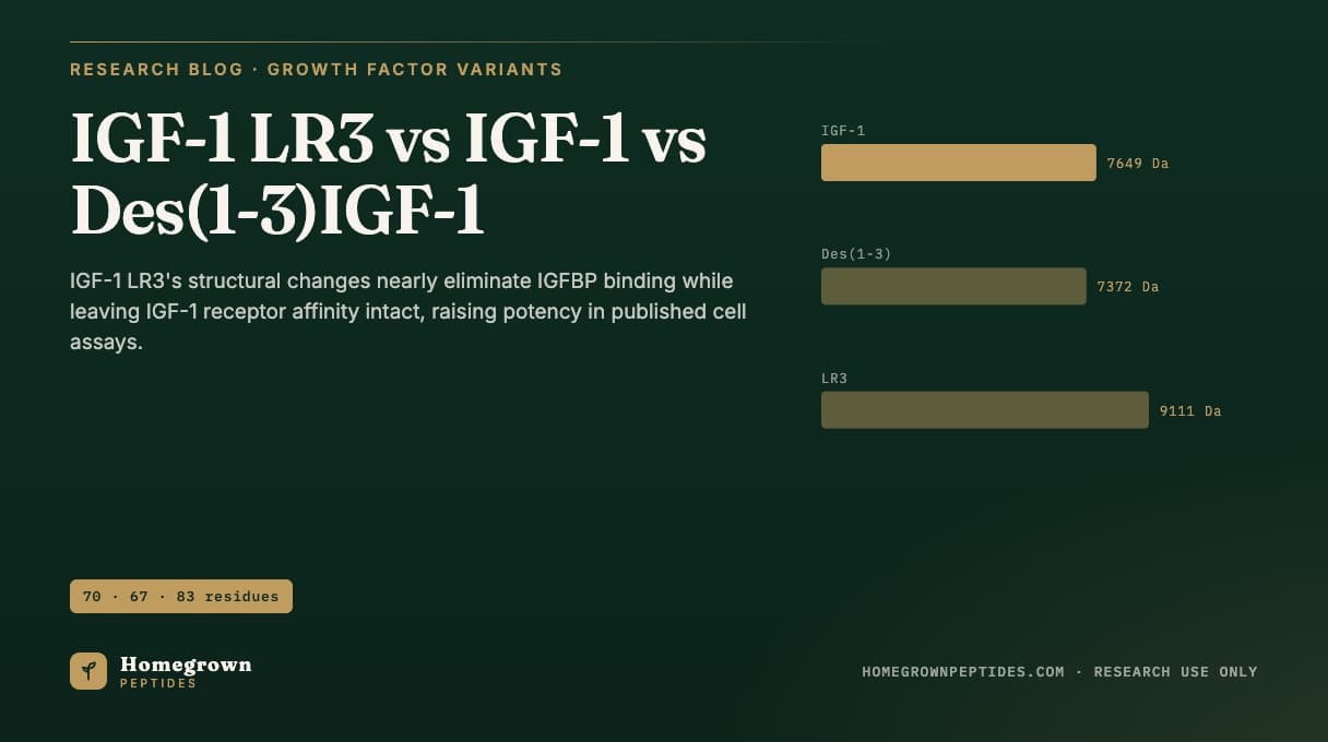
IGF-1 LR3 ("Long R3 IGF-1") is an 83-residue recombinant analog of human IGF-1 built by substituting arginine for the native glutamate at position 3 and fusing a 13-amino-acid extension peptide onto the N-terminus. Neither change touches the receptor-binding face of the molecule, but both sit in the region that insulin-like growth factor binding proteins (IGFBPs) use to grip IGF-1, so IGF-1 LR3 binds IGFBPs at roughly 1% or less of native IGF-1's affinity (Francis et al., 1992). In the fibroblast and L6 myoblast proliferation assays used to characterize it, that drop in binding-protein sequestration translated into two- to three-fold lower ED50 values than native IGF-1 at equivalent IGF-1 receptor affinity (Francis et al., 1992).
| Attribute | Detail |
|---|---|
| Native IGF-1 | 70 residues, ~7.65 kDa, full IGFBP and IGF-1R affinity (Ballard et al., 1987) |
| Des(1-3)IGF-1 | 67 residues (Gly-Pro-Glu removed from the N-terminus), ~7.4 kDa, markedly reduced IGFBP affinity (Francis et al., 1988) |
| IGF-1 LR3 | 83 residues (Arg3 substitution plus 13-residue N-terminal extension), ~9.1 kDa, IGFBP affinity reduced roughly 100-fold vs native IGF-1 (Francis et al., 1992) |
| Expression system reported | Recombinant fusion protein expressed in Escherichia coli, refolded and cleaved from the expression construct (Francis et al., 1992) |
| Identity/purity methods reported | SDS-PAGE, reversed-phase HPLC, amino acid sequencing/mass spectrometry, radioreceptor and cell-proliferation bioassay |
What does "LR3" mean in IGF-1 LR3?
LR3 stands for "Long R3": "Long" for the 13-amino-acid extension peptide fused onto the N-terminus, and "R3" for the arginine substituted at residue 3 in place of the native glutamate. Put together, the analog carries the complete 70-residue IGF-1 sequence with that single substitution, plus the extension, for a total of 83 residues. Francis and colleagues generated this and several related fusion-protein constructs specifically to probe how much of IGF-1's biological potency in cell assays is governed by IGF-1 receptor affinity versus sequestration by IGFBPs in the surrounding medium (Francis et al., 1992).
Why does the extension and Arg3 substitution reduce IGFBP binding?
IGFBPs recognize IGF-1 largely through contacts with its N-terminal region. Appending a 13-residue extension peptide and substituting the position-3 residue disrupts those contacts without touching the separate surface IGF-1 uses to engage its own receptor. That is the structural logic behind the result reported by Francis and colleagues: IGF-1 LR3 binds IGFBPs at a small fraction of native IGF-1's affinity while retaining IGF-1 receptor affinity comparable to the native hormone (Francis et al., 1992). Des(1-3)IGF-1 reaches a milder version of the same outcome by a different route, truncation rather than extension, removing the first three N-terminal residues (Gly-Pro-Glu) and reducing, though not eliminating, IGFBP binding (Francis et al., 1988).
How much more potent is IGF-1 LR3 in cell proliferation assays?
In the original characterization, Francis and colleagues tested these IGF-1 variants in fibroblast and L6 myoblast proliferation assays run in the presence of IGFBPs contributed by the culture medium or added back experimentally. Because IGF-1 LR3 is not effectively sequestered by IGFBPs in that assay system, more of the applied peptide remains free to engage IGF-1 receptors on the cells, which is reported as a lower ED50 (greater potency on a mass basis) relative to native IGF-1 run under the same conditions, even though direct IGF-1 receptor binding affinity for the two molecules is similar (Francis et al., 1992). The same logic, with a smaller effect size, explains why Des(1-3)IGF-1 also outperforms native IGF-1 in IGFBP-containing bioassays while showing receptor binding closer to native IGF-1 than to IGF-1 LR3 (Ballard et al., 1987; Francis et al., 1988).
Comparison: residues, affinity and assay potency
| Property | IGF-1 | Des(1-3)IGF-1 | IGF-1 LR3 |
|---|---|---|---|
| Residues | 70 | 67 | 83 |
| Structural change | None (native sequence) | N-terminal tripeptide removed | Arg3 substitution + 13-residue N-terminal extension |
| IGFBP affinity (relative to native IGF-1) | Reference (1x) | Markedly reduced (Francis et al., 1988) | Reduced roughly 100-fold (Francis et al., 1992) |
| IGF-1 receptor affinity | Reference (1x) | Close to native IGF-1 (Ballard et al., 1987) | Comparable to native IGF-1 (Francis et al., 1992) |
| Fibroblast/myoblast assay potency vs native IGF-1 | Reference | Several-fold more potent in IGFBP-containing medium | Two- to three-fold more potent in IGFBP-containing medium (Francis et al., 1992) |
| Expression system reported | Native/recombinant | Isolated from bovine colostrum; also produced recombinantly | Recombinant fusion protein, E. coli |
How is recombinant IGF-1 LR3 produced and characterized?
The published construct is expressed as a fusion protein in E. coli, then recovered, refolded and separated from expression-related fragments (Francis et al., 1992). Downstream characterization in this and related IGF-1 variant studies combines several orthogonal methods: SDS-PAGE to confirm apparent molecular weight and detect gross degradation or aggregation products, reversed-phase HPLC to resolve the intact peptide from truncated or oxidized species and estimate purity by peak area, and amino acid sequencing or mass spectrometry to confirm the expected sequence and mass. A cell-based bioassay, such as the fibroblast or myoblast proliferation assays used in the original characterization, or a radioreceptor binding assay against IGF-1 receptor preparations, is run alongside analytical testing because identity and purity data alone do not establish biological potency. For background on how mass spectrometry confirms peptide identity in general, see mass spectrometry for peptide identity, and for how reversed-phase HPLC purity numbers are generated and what they do and do not capture, see HPLC purity explained.
What do the published stability and storage notes say?
Ballard and colleagues, summarizing work on Des(1-3)IGF-1 and related truncated and fusion IGF-1 analogs, note that these molecules are handled using the same lyophilized-storage and reconstitution precautions applied to native IGF-1, since the modifications sit away from residues implicated in oxidative or deamidation-prone regions of the protein (Ballard et al., 1996). None of the characterization papers in this line of research report a stability profile distinct from native IGF-1 that would justify different handling. General peptide storage and freeze-thaw considerations that apply across this class of research reagents are covered in our IGF-1 LR3 reference page, which also summarizes lot-specific analytical expectations.
Does IGF-1 LR3 cross-react with the insulin receptor?
Native IGF-1 has long been reported to bind the insulin receptor with much lower affinity than insulin binds its own receptor, a cross-reactivity relationship driven by sequence and structural similarity between the IGF-1 and insulin receptor families. Mapping work on the IGF-1 receptor binding surface has shown that the residues governing receptor engagement are distinct from the N-terminal region IGFBPs use for recognition (Keyhanfar et al., 2007), which is consistent with why neither the Des(1-3)IGF-1 truncation nor the IGF-1 LR3 extension and Arg3 substitution is reported to shift insulin-receptor cross-reactivity in a way that distinguishes these analogs from native IGF-1 in the original characterization studies (Francis et al., 1992). In practice, insulin receptor cross-reactivity for all three molecules is treated in the literature as a feature of the conserved IGF-1 receptor-binding surface, not of the N-terminal region that differs between them.
What has this comparison been examined in, in animal models?
Outside cultured cells, IGF-1 variants with reduced IGFBP affinity have also been examined in whole-animal studies. Tomas and colleagues compared native IGF-1 with an IGF-1 variant in normal female rats and reported differences in growth and nitrogen-retention measures consistent with the variant circulating less bound to IGFBPs and more available to engage receptors in peripheral tissue, paralleling the cell-assay potency differences reported by Francis and colleagues (Tomas et al., 1993). This body of work, taken together, treats IGFBP affinity as the main variable separating these three molecules' behavior in both cultured cells and intact animals, with IGF-1 receptor affinity held roughly constant across all three (Tomas et al., 1993; Francis et al., 1992).
A broader account of IGF-1 LR3's origin, structure and the IGFBP literature behind it is covered in what is IGF-1 LR3; this article focuses specifically on how its binding-protein affinity and cell-assay potency numbers compare against native IGF-1 and Des(1-3)IGF-1 side by side. Lot-specific identity and purity data for research-grade material are posted on our IGF-1 LR3 lot page.
Frequently asked questions
Is IGF-1 LR3 the same molecule as Des(1-3)IGF-1?
No. Des(1-3)IGF-1 is native IGF-1 with the first three N-terminal residues removed, giving a 67-residue molecule. IGF-1 LR3 is a larger, 83-residue fusion construct carrying the full native IGF-1 sequence, an Arg3 substitution, and a 13-residue N-terminal extension. Both reduce IGFBP binding relative to native IGF-1, but through different structural changes and to different degrees.
Does IGF-1 LR3 bind the IGF-1 receptor more tightly than native IGF-1?
Published characterization reports IGF-1 LR3's IGF-1 receptor affinity as comparable to, not substantially higher than, native IGF-1. The potency difference seen in cell assays is attributed mainly to reduced IGFBP sequestration, not to improved receptor binding (Francis et al., 1992).
What cell types were used in the original potency comparisons?
The characterization papers in this line of research used fibroblast cultures and L6 myoblast proliferation assays, run in the presence of IGFBPs contributed by the culture system, to compare potency across native IGF-1, Des(1-3)IGF-1 and IGF-1 LR3 (Francis et al., 1992).
How is IGF-1 LR3 identity typically confirmed on a certificate of analysis?
Common methods reported in the literature and used on commercial certificates include SDS-PAGE for apparent molecular weight, reversed-phase HPLC for purity, and mass spectrometry or amino acid sequencing for sequence confirmation.
Has IGF-1 LR3 been studied in humans?
The characterization and comparison literature cited here consists of cell culture and rodent studies. This article summarizes laboratory research models, not clinical evidence, and nothing here describes a protocol for use in any subject.
Why does reduced IGFBP binding matter for a cell assay?
IGFBPs in culture medium or serum sequester IGF-1, lowering the amount of free peptide available to engage cell-surface receptors. A molecule that binds IGFBPs weakly stays more available to the receptor at the same applied concentration, which is reported as a lower ED50 in proliferation assays (Francis et al., 1992).
Is Des(1-3)IGF-1 found naturally, or is it only made synthetically?
It was first characterized from bovine colostrum as a naturally occurring truncated form of IGF-1, and has also been produced recombinantly for research use (Francis et al., 1988).
References
- Ballard FJ, Francis GL, Ross M, Bagley CJ, May B, Wallace JC. Natural and synthetic forms of insulin-like growth factor-1 (IGF-1) and the potent derivative, destripeptide IGF-1: biological activities and receptor binding. Biochem Biophys Res Commun. 1987;149(2):398-404. PubMed 2962574.
- Francis GL, Upton FM, Ballard FJ, McNeil KA, Wallace JC. Insulin-like growth factors 1 and 2 in bovine colostrum. Sequences and biological activities compared with those of a potent truncated form. Biochem J. 1988;251(1):95-103. PubMed 3390164.
- Francis GL, Ross M, Ballard FJ, Milner SJ, Senn C, McNeil KA, Wallace JC, King R, Wells JR. Novel recombinant fusion protein analogues of insulin-like growth factor (IGF)-I indicate the relative importance of IGF-binding protein and receptor binding for enhanced biological potency. J Mol Endocrinol. 1992;8(3):213-23. PubMed 1378742.
- Tomas FM, Knowles SE, Chandler CS, Francis GL, Owens PC, Ballard FJ. Anabolic effects of insulin-like growth factor-I (IGF-I) and an IGF-I variant in normal female rats. J Endocrinol. 1993;137(3):413-21. PubMed 8371075.
- Ballard FJ, Wallace JC, Francis GL, Read LC, Tomas FM. Des(1-3)IGF-I: a truncated form of insulin-like growth factor-I. Int J Biochem Cell Biol. 1996;28(10):1085-7. PubMed 8930132.
- Keyhanfar M, Booker GW, Whittaker J, Wallace JC, Forbes BE. Precise mapping of an IGF-I-binding site on the IGF-1R. Biochem J. 2007;401(1):269-77. PubMed 16981855.
- Clemmons DR. Insulin-like growth factor binding proteins: roles in regulating IGF physiology. J Dev Physiol. 1991;15(2):105-10. PubMed 1713934.
Research use only. Homegrown Peptides products are for laboratory research and are not for human or animal use. Nothing in this article is medical advice.