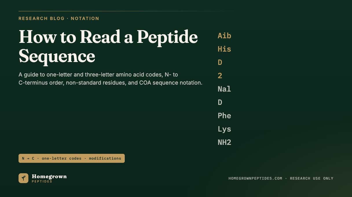
A peptide sequence is written left to right from the N-terminus (free amine end) to the C-terminus (free carboxyl end), using one-letter or three-letter codes for each amino acid. Everything else attached to that string, a prefix like Ac-, a suffix like -NH2, a D- residue like Aib, a bracket linking two cysteines, tells a reader what was changed from the plain chain, and each change shifts the mass by a calculable amount.
| What you see | What it means |
|---|---|
| Left-to-right order | N-terminus first, C-terminus last, per IUPAC-IUB nomenclature |
| Capital single letter (G, K, F) | Standard L-amino acid, one of 20 genetically coded residues |
| Lowercase letter or "D-" prefix | D-stereoisomer; same formula and mass as the L-form, different shape |
| Ac- prefix | N-terminal acetylation, adds about 42.01 Da monoisotopic |
| -NH2 suffix | C-terminal amidation, subtracts about 0.98 Da monoisotopic |
| Non-standard code (Aib, Nle, Orn, Nal) | Residue outside the 20 standard, with its own fixed formula |
| Two Cys linked by a bracket | Disulfide bond connecting those side chains, cyclizing the backbone |
What is the correct direction to read a peptide sequence?
Every peptide chain has two chemically distinct ends: an N-terminus with a free amine, and a C-terminus with a free carboxyl, unless either end is capped. The 1983 IUPAC-IUB nomenclature recommendations fixed the convention that a sequence is written and numbered N-terminus to C-terminus, matching the direction a ribosome synthesizes a chain. In "His-Trp-Ser-Tyr", His is residue 1 with the free amine and Tyr is last with the free carboxyl; reversing the order describes a different molecule, even with the same four amino acids.
One-letter versus three-letter codes: when does each get used?
Three-letter codes (Gly, Ala, Lys) are easier to read at a glance and standard in prose and most COA headers. One-letter codes (G, A, K) pack a sequence into a compact string and are the default in sequence databases, alignment tools and FASTA files. Three ambiguity codes exist for cases a method cannot resolve: B for Asx (Asn or Asp), Z for Glx (Gln or Glu), and X for any unidentified residue. A COA for a well-characterized short research peptide should never need these; they belong in de novo sequencing of unknowns.
| Amino acid | 3-letter | 1-letter |
|---|---|---|
| Alanine | Ala | A |
| Arginine | Arg | R |
| Asparagine | Asn | N |
| Aspartic acid | Asp | D |
| Cysteine | Cys | C |
| Glutamic acid | Glu | E |
| Glutamine | Gln | Q |
| Glycine | Gly | G |
| Histidine | His | H |
| Isoleucine | Ile | I |
| Leucine | Leu | L |
| Lysine | Lys | K |
| Methionine | Met | M |
| Phenylalanine | Phe | F |
| Proline | Pro | P |
| Serine | Ser | S |
| Threonine | Thr | T |
| Tryptophan | Trp | W |
| Tyrosine | Tyr | Y |
| Valine | Val | V |
What residues show up in catalog peptides that are not on that list?
Research peptides often depart deliberately from the 20 standard residues: a non-standard residue can block the enzyme that would otherwise cut the chain, or lock a backbone angle a flexible side chain cannot hold. Spyroulias and colleagues showed this directly, swapping a Phe for D-Phe and a Leu for Aib in corticotropin releasing hormone and tracking how each substitution changed the local structure (Spyroulias et al., 2002).
| Residue | What it is | Why used |
|---|---|---|
| Aib (α-aminoisobutyric acid) | Alanine with a second methyl on the alpha carbon | Rigid helical turn, resists aminopeptidase cleavage |
| Nle (norleucine) | Straight-chain isomer of leucine/isoleucine | Avoids methionine's oxidation-prone side chain |
| D-Ala, D-Phe, D-2-Nal, other D-residues | Mirror-image stereoisomers of L-amino acids; same formula and mass | Changes receptor fit, resists stereospecific proteases |
| Orn (ornithine) | Lysine homolog, one methylene shorter | Used in some bioregulator and cyclic-peptide designs |
| 2-Nal (2-naphthylalanine) | Phenylalanine with a two-ring naphthyl group | Bulkier, more hydrophobic aromatic contact point |
| Pal (palmitoyl) on Lys | C16 fatty acyl group amide-bonded to a lysine side chain | Anchors to circulating albumin in long-acting incretin analogs |
| γ-Glu linker | Glutamic acid joined through its side-chain carboxyl | Flexible spacer between a lysine side chain and a fatty acid |
| AEEA / OEG spacer | 8-amino-3,6-dioxaoctanoic acid, a PEG-like unit | Adds distance and hydrophilicity before a lipid group |
These appear written inline (Aib-His-D-2-Nal-D-Phe-Lys-NH2) or, for heavily modified molecules, as shorthand against a parent sequence, such as "Aib8, Arg34, Lys26(γGlu-γGlu-C18 diacid)-GLP-1(7-37)" for a fatty-acid-conjugated incretin analog. D-residue substitutions also blunt enzymatic cleavage in cyclic peptides, as shown for a conotoxin where swapping single D-residues identified which positions controlled potency and proteolysis resistance (Ren et al., 2019).
How are terminal modifications written, and what do they do to the mass?
A plain sequence like "His-Trp-Ser-Tyr" implies a free amine at the N-terminus and a free acid at the C-terminus. Capping either end is common and always flagged in the notation. Ac- (acetyl) is the most common N-terminal cap, adding roughly 42.01 Da monoisotopic. pGlu- (pyroglutamate) is a cyclized Gln or Glu at position 1 that loses water (about -18.01 Da) relative to the open chain. -NH2 (amide) is the most common C-terminal cap in short research peptides, subtracting about 0.98 Da monoisotopic from the free-acid mass; a free acid is the default unless a cap is noted. Both caps block the exopeptidases that chew in from a free terminus. The full math behind residue masses and water loss at each bond is worked step by step in how to calculate a peptide's molecular weight and formula from its sequence.
How is a disulfide bond or cyclic backbone shown in a sequence?
A linear string of letters cannot by itself show a bond between two side chains that are not adjacent in the chain, so notation adds a separate annotation. The common convention numbers the cysteines and states which pair is bridged, for example "Cys-Tyr-Phe-Gln-Asn-Cys-Pro-Arg-Gly-NH2, disulfide bond Cys1-Cys6" for a ring closed between the first and sixth residues; papers and databases sometimes draw this as a bracket instead. A disulfide bridge is a real structural constraint, not a cosmetic label: breaking it changes the mass by exactly 2.02 Da (two hydrogens) relative to the reduced, linear form, which a mass-spec check must account for. Work redesigning the disulfide ring systems of oxytocin and vasopressin analogs shows how changing which cysteines are bridged shifts stability and selectivity while the linear sequence stays the same (Beard et al., 2018). Backbone cyclization, joining the N- and C-termini instead of two side chains, is noted as "cyclo(" before the sequence and ")" after it.
Worked example: reading and calculating ipamorelin's sequence
Ipamorelin's published sequence is Aib-His-D-2-Nal-D-Phe-Lys-NH2, a pentapeptide (Raun et al., 1998). Reading left to right: Aib sits at the N-terminal position, His and Lys are standard L-residues, D-2-Nal and D-Phe are D-stereoisomers, and the chain ends in an amide rather than a free acid.
Step 1, residue formulas. Aib = C4H7NO, His = C6H7N3O, D-2-Nal = C13H11NO, D-Phe = C9H9NO, Lys = C6H12N2O. D- and L-residues share the same formula.
Step 2, sum, add water, apply the amide. Summing elements across the five residues plus one H2O gives a free-acid formula of C38H48N8O6. Amidation then replaces -OH with -NH2 (remove one O, add one N and one H), giving C38H49N9O5.
Step 3, sum the masses. Standard monoisotopic residue masses (Aib 85.0528, His 137.0589, D-2-Nal 197.0841, D-Phe 147.0684, Lys 128.0949) sum to 694.3591 Da; adding one water gives 712.3697 Da free acid, and the amide shift of -0.9840 Da brings the final mass to approximately 711.39 Da monoisotopic, near 711.86 Da average. A lot's certificate should report an observed mass within tolerance; a bigger gap points to the lot, not the arithmetic.
For comparison, semax, Met-Glu-His-Phe-Pro-Gly-Pro, is a heptapeptide of standard L-residues with no caps (Dolotov et al., 2006): nothing to track beyond the plain residue sum, while ipamorelin carries one non-standard residue, two D-stereocenters, and a C-terminal amide. Lot-specific identity data for both sit on their product pages: research-grade ipamorelin and research-grade semax.
How do sequence databases like UniProt and PubChem present a sequence?
UniProt, the main curated protein sequence database, displays entries in FASTA format: a header line starting with ">" followed by an unbroken one-letter-code string, with no spaces or modification symbols in the letters themselves (Zaru et al., 2023). Any modification, a cleaved signal peptide, a disulfide bond, a glycosylation site, is recorded as a separate annotation on a specific residue position, not inline in the string. That fits full-length proteins built from the 20 standard residues, most of what UniProt catalogs.
PubChem, by contrast, is built around small molecules and short peptides and represents a compound by its structural formula and machine-readable connectivity strings (SMILES, InChI) rather than a letter sequence (Kim et al., 2023). A peptide carrying Aib, a D-residue, or a lipid-conjugated lysine is usually easier to look up correctly in PubChem by structure or exact mass than to force into a letter sequence never designed to carry those symbols.
How does the sequence line on a certificate of analysis tie back to the mass spec check?
A certificate of analysis states the sequence as text, alongside a target molecular formula and mass, then reports what the LC-MS run measured. The instrument never reads letters; it measures mass-to-charge ratios for charged ions, and a deconvolution step converts that into a single neutral mass, the same kind of intact-mass measurement used to confirm subunit identity in other characterized proteins (Rinalducci et al., 2009). That observed mass is compared against the mass calculated from the stated sequence, residue by residue, with every cap, substitution, and disulfide accounted for. Agreement within tolerance supports that the vial contains the labeled sequence; it does not confirm stereochemistry at a D-residue, since D- and L-forms are indistinguishable by mass alone. See mass spectrometry for peptide identity for how that matching works, and how to read a peptide certificate of analysis for where the sequence line sits relative to purity and endotoxin results. Counter-ion identity, whether a basic peptide is a trifluoroacetate or acetate salt, is reported separately and never appears in the sequence notation; see TFA vs acetate salt forms of research peptides.
Frequently asked questions
Is a peptide sequence ever written C-terminus to N-terminus?
By the IUPAC-IUB convention, no. Standard practice is always N-terminus first, C-terminus last. A source that writes it the other way without saying so has a labeling error, not an accepted convention.
Does a lowercase letter always mean a D-amino acid?
Not universally. Many sources mark D-residues with an explicit "D-" prefix (D-Phe, D-Ala) and use lowercase only as a typographic choice elsewhere. Check how a given source defines its own notation before assuming.
Why do some sequences list only a few residues with the rest implied?
For peptides derived from a well-known parent, such as a GLP-1 or GHRH analog, papers often write only the positions that differ from the parent, with the parent sequence assumed.
Can two peptides have the same amino acid composition but different sequences?
Yes. The same residues in a different order form a different molecule with identical formula and mass but different chemistry and activity. Plain mass measurement cannot distinguish sequence isomers; that requires MS/MS fragmentation or a sequencing method that reads residue order directly.
What does an "X" in a sequence mean?
X denotes a position a sequencing method could not assign. A commercial research peptide's labeled sequence should not contain one; X belongs in de novo or unknown-sample sequencing, not a defined synthetic product.
References
- Kim S, Chen J, Cheng T, et al. PubChem 2023 update. Nucleic Acids Res. 2023;51(D1):D1373-D1380. PMID 36305812.
- Zaru R, Orchard S, UniProt Consortium. UniProt Tools: BLAST, Align, Peptide Search, and ID Mapping. Curr Protoc. 2023;3(3):e697. PMID 36943033.
- Raun K, Hansen BS, Johansen NL, et al. Ipamorelin, the first selective growth hormone secretagogue. Eur J Endocrinol. 1998;139(5):552-561. PMID 9849822.
- Dolotov OV, Karpenko EA, Inozemtseva LS, et al. Semax, an analog of ACTH(4-10) with cognitive effects, regulates BDNF and trkB expression in the rat hippocampus. Brain Res. 2006;1117(1):54-60. PMID 16996037.
- Spyroulias GA, Nikolakopoulou P, Tzakos A, et al. Monitoring the structural consequences of Phe12-D-Phe and Leu15-Aib substitution in human/rat corticotropin releasing hormone. Eur J Biochem. 2002;269(24):6009-6019. PMID 12473096.
- Ren J, Yu Z, Chen G, et al. D-Amino Acid Substitution of alpha-Conotoxin RgIA Identifies its Critical Residues and Improves the Enzymatic Stability. Mar Drugs. 2019;17(3):142. PMID 30823399.
- Beard R, Stucki A, Schmid M, et al. Building bridges for highly selective, potent and stable oxytocin and vasopressin analogs. Bioorg Med Chem. 2018;26(11):3039-3045. PMID 29602673.
- Rinalducci S, Roepstorff P, Zolla L. De novo sequence analysis and intact mass measurements for characterization of phycocyanin subunit isoforms from the blue-green alga Aphanizomenon flos-aquae. J Mass Spectrom. 2009;44(4):503-515. PMID 19053161.
Research use only. Homegrown Peptides products are for laboratory research and are not for human or animal use. Nothing in this article is medical advice.